
What an OSCE station is
- Door chart Read before you knock
- History, 7 min Ask what matters most first.
- Physical exam, 5 min A focused exam, with findings as you go.
- Assessment and plan, 3 min Your differential and next steps, explained to the patient.
In an Objective Structured Clinical Examination (OSCE), you rotate through a series of stations. At each one you complete a defined task or set of tasks, such as taking a history, performing part of the exam or interpreting results, and examiners often score it with a checklist written in advance.1,2
The format is now used widely in undergraduate and postgraduate clinical education.2 The details are not fixed: each program decides how many stations to run, how long they last, what each asks of you and how it is scored.3 Read your own program's instructions before you rely on any single description, including this one.
This sample uses a common format: a door chart you read before you go in, then 15 minutes with the patient, then a short presentation to the examiner. The pace in the figure is a suggestion for this case, not a rule.
How the station goes
The patient is Maya Collins, a 24-year-old woman with abdominal pain, and you have 15 minutes. One moment from each stage shows what it feels like, with what an examiner credits there and the miss that costs the most points.
-
Before you knock
Read the door chart
Maya Collins, 24. Abdominal pain since last night. Temp 38.1 °C (100.6 °F), heart rate 102/min.
Read the vital signs, not just the complaint, and write a short differential before you knock.
Common miss Skipping the vitals: her fever and fast heart rate make this more than a stomachache.
-
About 7 minutes
Take a focused history
You Where is the pain, and has it moved since it started?
Maya It started around my belly button. Sometime this morning it moved down here.
13 items on the examiner's checklist.
Common miss Leaving the menstrual and sexual history for last and running out of time.
-
About 5 minutes
Examine the patient
You Palpate all four quadrants, starting on the left.
Finding Soft. Tender in the right lower quadrant, most at McBurney point, with voluntary guarding. The other quadrants are not tender.
9 items on the examiner's checklist, including the Rovsing, psoas and obturator signs.
Common miss Spending minutes on maneuvers that have nothing to do with abdominal pain.
-
Part of the last 3 minutes
Order the workup
You Order a serum pregnancy test (hCG) first.
Result Negative, below 5 mIU/mL. Then CT: dilated appendix, 11 mm, with wall thickening and surrounding fat stranding.
4 items on the examiner's checklist.
Common miss Ordering a plain abdominal x-ray. Not the study of choice for right lower quadrant pain. CT with IV contrast is.
-
Part of the last 3 minutes
Give your assessment and plan
You The scan shows that your appendix is inflamed. That's appendicitis. It's usually treated with surgery, so I've asked the surgeons to see you.
Maya Okay. Will I still make my interview on Friday?
6 items on the examiner's checklist: the leading diagnosis, the diagnoses you cannot miss, and a safe plan.
Common miss Explaining it in clinical shorthand. The content is right, but the patient cannot follow it, and it does not check that she understood.
-
After the encounter
Present the case
You My assessment is acute appendicitis without signs of perforation.
Present in four short parts: the story, the exam, the results, then your assessment and plan.
Common miss Hedging. “It could be a lot of things” is not an assessment.
History checklist
The examiner credits each item yes or no. This is the sample station's list; yours will differ.
- Onset and duration. Timing frames the differential. Appendicitis usually comes on over hours, within about a day.
- Location and migration. Pain that starts around the umbilicus and moves to the right lower quadrant is one of the more useful history clues to appendicitis.
- Character and severity. How the pain has changed, and how bad it is now, sets a baseline to reassess against.
- Aggravating and relieving factors. Pain with movement, or with every bump in the road, suggests peritoneal irritation.
- Appetite. Loss of appetite is common in appendicitis, though on its own it is a weak clue.
- Nausea and vomiting, with timing. Nausea and vomiting are common in appendicitis and in many of its mimics, so ask about both and about their timing.
- Fever and chills. Fever supports an inflammatory or infectious cause.
- Bowel habits. Diarrhea would make gastroenteritis more likely, and a change in bowel habits matters for obstruction.
- Urinary symptoms. Urinary symptoms point toward infection or a stone.
- Last menstrual period. A period that is due or late means pregnancy cannot be ruled out by history.
- Sexual history and pregnancy risk. Sexual history shapes the risk of ectopic pregnancy and pelvic inflammatory disease.
- Past medical and surgical history. A prior appendectomy would change the differential.
- Medicines and allergies. You need both before you order contrast or give pain medicine.
Communication
Not checklist items. A real station might rate these on a 1 to 5 scale.
- Introduced self, confirmed the patient and opened with an open question
- Asked what worries her most
- Summarized what she said and checked for anything missed
Physical exam checklist
The examiner credits each item yes or no. This is the sample station's list; yours will differ.
- Inspects the abdomen. Distension, scars and how the patient lies all carry information.
- Listens to bowel sounds. Part of this sample's conventional exam sequence, though bowel sounds play a limited role in diagnosis.
- Palpates all four quadrants. Palpating every quadrant, starting away from the pain, localizes the tenderness.
- Checks for peritoneal irritation. Percussion tenderness or rebound tenderness shows peritoneal irritation. Percussion is the gentler test, and one positive sign is enough.
- Rovsing sign. Each special maneuver has low sensitivity, but a positive one modestly raises the likelihood of appendicitis.
- Psoas sign. A positive psoas sign suggests an inflamed appendix lying behind the cecum.
- Obturator sign. A positive obturator sign suggests a pelvic appendix. A negative one does not rule appendicitis out.
- Costovertebral angle tenderness. Flank tenderness would point toward the kidney.
- Pelvic exam. In a woman with lower abdominal pain, the pelvic exam looks for cervical motion tenderness and adnexal masses.
Communication
Not checklist items. A real station might rate these on a 1 to 5 scale.
- Explained the exam and kept her draped
Workup checklist
The examiner credits each item yes or no. This is the sample station's list; yours will differ.
- Pregnancy test. Every premenopausal woman with acute abdominal pain needs one. It decides the imaging, and a positive result calls for pelvic ultrasound to find where the pregnancy is.
- Blood count and chemistry. Part of the standard workup for acute abdominal pain.
- Urinalysis. Looks for infection or blood that would point to a urinary cause.
- CT with IV contrast. The imaging test of choice for right lower quadrant pain in a patient who is not pregnant.
Assessment and plan checklist
The examiner credits each item yes or no. This is the sample station's list; yours will differ.
- Leading diagnosis: acute appendicitis. Migration to McBurney point, anorexia, fever and peritoneal signs make it the leading diagnosis.
- Considers ectopic pregnancy. In a woman who could be pregnant, ectopic pregnancy is the diagnosis you cannot miss.
- Considers another gynecologic cause. Adnexal torsion and pelvic inflammatory disease belong on the differential for lower abdominal pain in a woman.
- Nothing by mouth, IV fluids. She may need surgery, and she has been vomiting.
- Pain control. Pain medicine should not be withheld; it does not make the exam less accurate.
- Surgical consultation. Consult early when appendicitis is likely: delay is associated with perforation.
Communication
Not checklist items. A real station might rate these on a 1 to 5 scale.
- Explained the diagnosis and plan in plain language and invited questions
How OSCE stations are scored
A station can be scored with a checklist, a rating scale, or both.3,4 A checklist lists specific actions, each credited yes or no: asked about the last menstrual period, checked for peritoneal irritation, ordered a pregnancy test. Because the items are written before the exam, every student is marked against the same list.1
A global rating summarizes the quality of a broad performance domain, such as communication or organization, or of the encounter overall, on a scale. In studies using expert examiners, global ratings were at least as reliable as checklists and better at separating experienced clinicians from trainees.5,6 A checklist counts what you did; a rating weighs how well you did it.
This sample's checklist has 32 items in three domains. A real station might add an examiner's 1 to 5 rating of communication, based on behaviors such as introducing yourself, asking about the patient's concerns and explaining the plan in plain language.
Working with a standardized patient
A standardized patient (SP) is a person trained to present an illness in a standardized way: usually a well person portraying a case, sometimes an actual patient presenting their own illness.7
- The same case for everyone SPs are trained to portray a case realistically and repeatably, so every student meets the same case facts.4 How tightly the wording is scripted depends on the purpose of the encounter.
- Ask for what you need Don't count on the patient to volunteer the clue that matters. In this sample, Maya answers only what you ask, and nobody mentions the period that is due unless you ask about it.
- Findings that are not in the room Some findings, like a fever, cannot be portrayed. Stations handle this in different ways; this sample gives you a findings card from the examiner.
The clinical reasoning behind this case
Where the pain is narrows the list
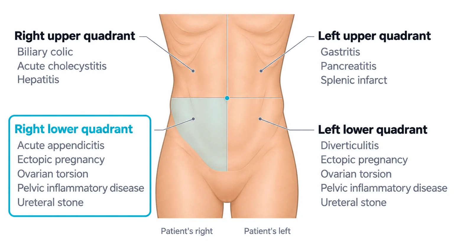
The location of the pain is the first filter for acute abdominal pain.8 Right lower quadrant pain in a woman of reproductive age has surgical, gynecologic and urinary causes, so the differential starts wide: appendicitis, ectopic pregnancy, ovarian torsion, pelvic inflammatory disease and a ureteral stone.
Keep the gynecologic causes on the list at least until the pregnancy test and the pelvic exam are back, and longer if the findings point that way.8
What points to appendicitis
Maya's story is a common presentation of acute appendicitis, which usually presents within about 24 hours, though the course varies and a longer history does not rule it out.9 Right lower quadrant pain is one of the strongest findings, and pain that began near the umbilicus and moved there adds to it. Loss of appetite and vomiting are common but weak clues on their own.10
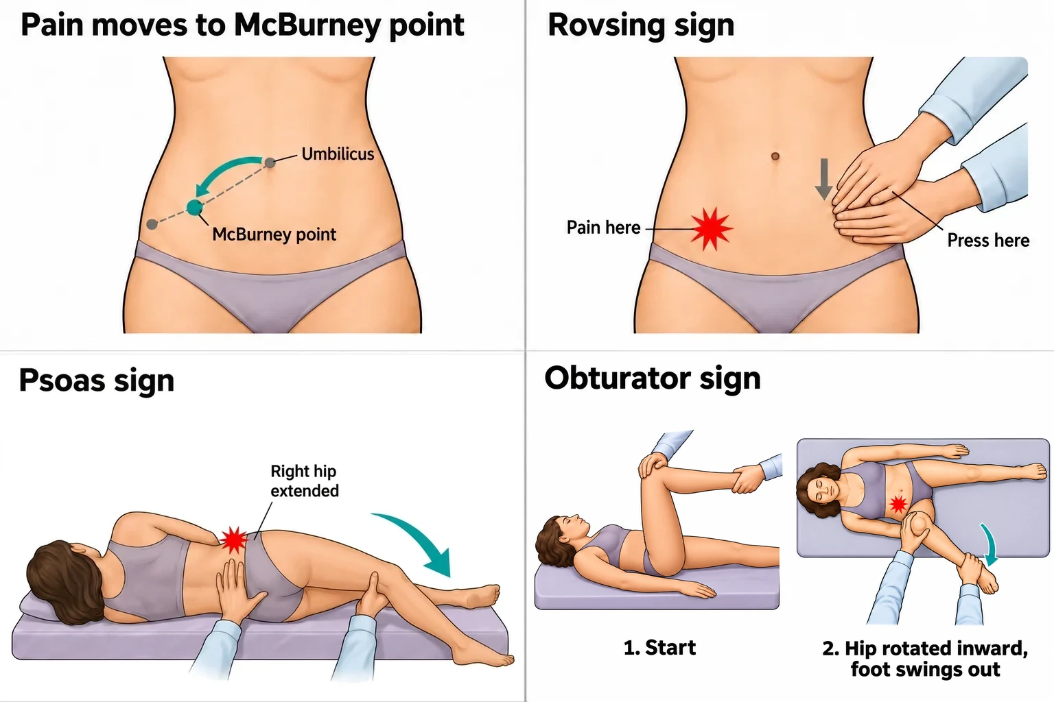
Tenderness is classically greatest at McBurney point, about one third of the way along the line from the right anterior superior iliac spine to the umbilicus.11 Percussion tenderness and rebound tenderness signal peritoneal irritation; percussion is the gentler test.8
Rovsing sign is right lower quadrant pain when you press deeply on the left lower quadrant; the psoas sign is pain when the right hip is extended with the patient on her left side; the obturator sign is pain on internal rotation of the flexed right hip.12 These signs have low sensitivity but modestly raise the likelihood of appendicitis when present,8 so Maya's negative obturator sign does not rule it out.
What argues against the others
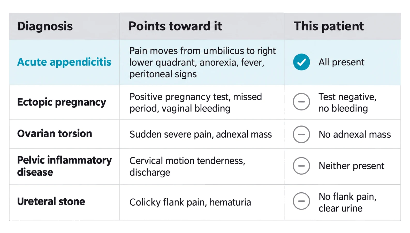
Each diagnosis that stays on the list has a finding that would point toward it. Ectopic pregnancy needs a pregnancy; a missed period or vaginal bleeding raises concern.8 Ovarian torsion often presents with nonspecific lower abdominal pain, sometimes with nausea and vomiting.8 The pain is classically sudden in onset, and an adnexal mass is common, but torsion can occur without one, so a normal pelvic exam does not exclude it.13 Pelvic inflammatory disease is suggested by cervical motion, uterine or adnexal tenderness.14 A ureteral stone often causes colicky flank pain and may cause blood in the urine, though its absence does not exclude a stone.8
Maya's pregnancy test is negative, and she has no vaginal bleeding, adnexal mass, cervical motion tenderness, flank pain or blood in the urine. That is why a strong student can rank appendicitis first and say why each of the others is less likely.
The pregnancy test comes first
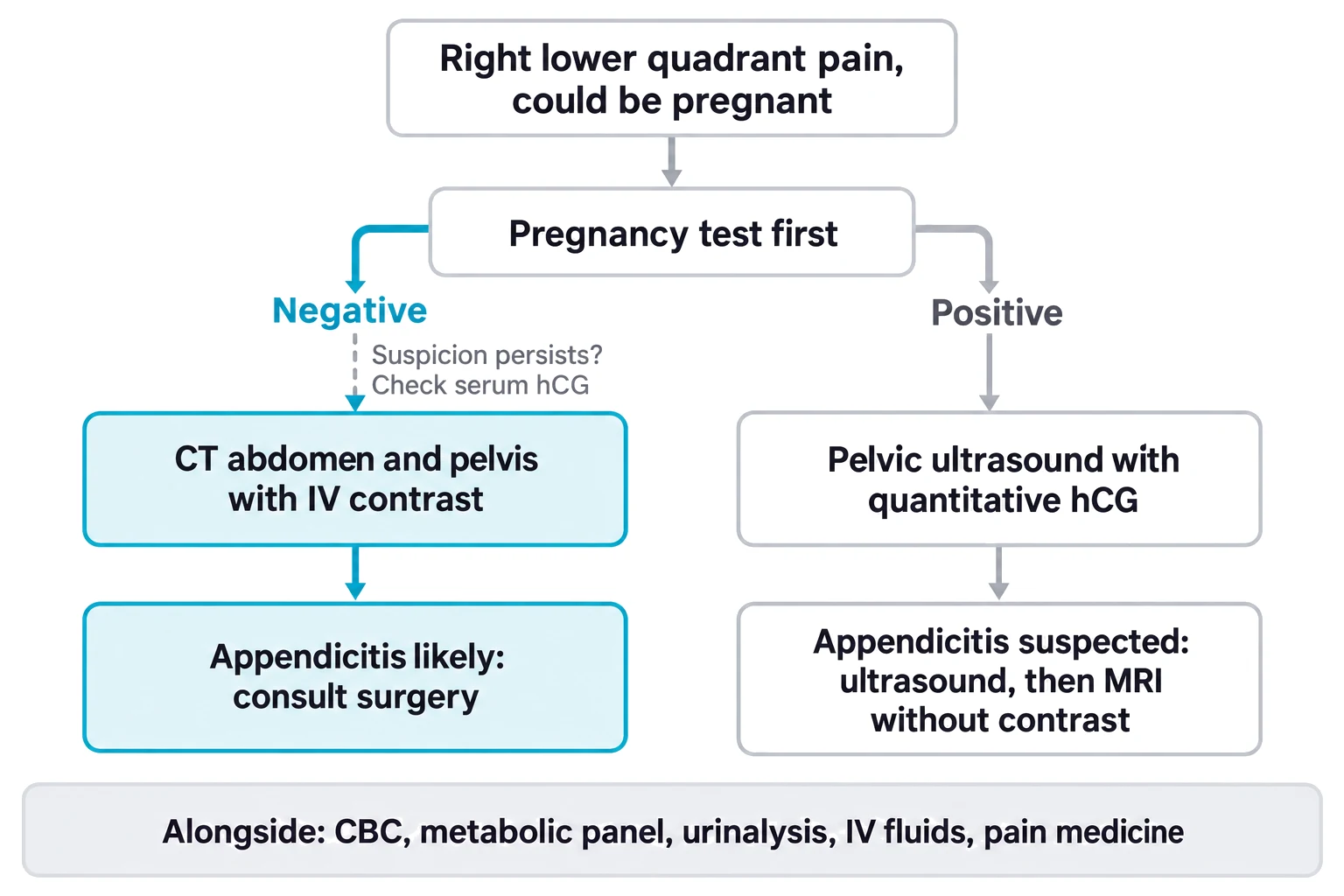
Order a pregnancy test in every premenopausal woman with acute abdominal pain.8 The test shows whether she is pregnant, not where. A negative urine test can miss an early pregnancy, ectopic pregnancies included,15 so check a serum level when suspicion persists. A positive test calls for pelvic ultrasound to look for an intrauterine pregnancy, read with a quantitative serum hCG.16
If she is not pregnant and appendicitis leads the list, CT of the abdomen and pelvis with IV contrast is the imaging test of choice; in pregnancy, ultrasound and MRI without contrast come first.8,17 Give pain medicine, which does not make the exam less accurate.8,10 When appendicitis is likely, consult surgery early.10 Treatment, appendectomy or, in selected uncomplicated cases, antibiotics alone, is decided with the surgical team.10,18
How to practice for an OSCE
- Say it out loud, on a clock Practice with a classmate playing the patient, or alone, speaking every question. Time it the way your program times its stations.
- Build a frame for each complaint For each common chief complaint, know the questions that change the workup and ask them early. Here: migration, appetite, fever, and the menstrual and sexual history.
- Rehearse the exam as a sequence Run the maneuvers in the same order until it is automatic, and say what you are doing and what you find. Follow your program's instructions about verbalizing findings.
- Debrief on content and communication After each attempt, score yourself on what you did and on how you explained it, then repeat the case or a similar one.